 Lasers & Optical Fibers Technologies
RMC2.5 Re-scan Confocal Microscope VIS-NIR / Microscopes imaging
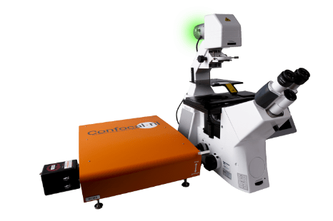
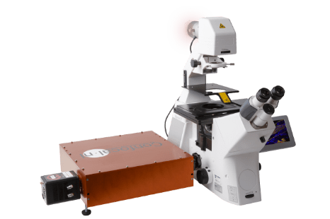
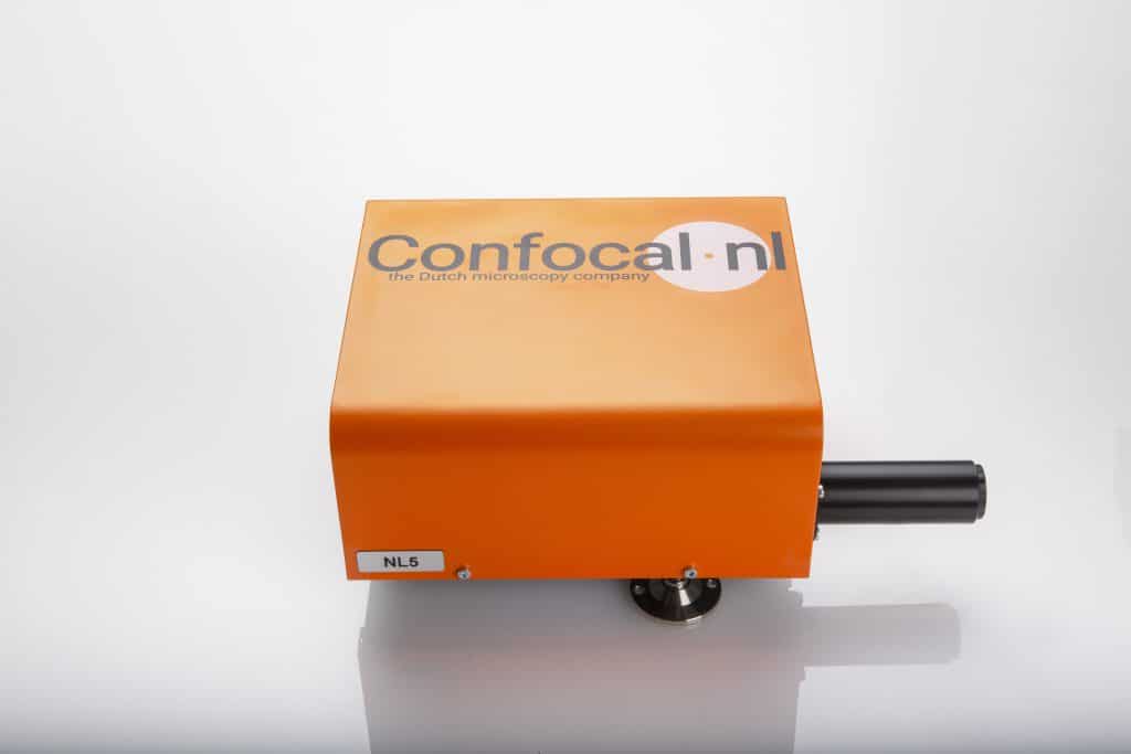
The RCM2.5 is the next big thing in confocal microscopy. Building on the existing RCM2 platform, we have developed a version that can utilize up to 5 lasers; extending over the Visible and Near-Infrared (NIR). Moreover, RCM2.5 enables you to use the latest advances in NIR dye development and look much deeper into your specimen. Experience complete experimental freedom and flexibility with our RCM2.5

RMC2.5 Re-scan Confocal Microscope VIS-NIR summary
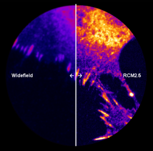
With a 5th channel, you can expand your experiment with an extra label, enabling you to see more details and gather even more relevant information. The NIR window allows for deeper imaging in biological specimens. Our re-scanning technology improves the lateral resolution of the microscope also at 785 nm, allowing higher resolution imaging compared to traditional confocal microscopes. Moreover, the camera-based detection in the RCM has a much higher efficiency compared to PMTs. With RCM2.5, you’ll work more efficiently, save time, and most importantly: produce high contrast images of the best image quality you can imagine. Acquiring fluorescent images has never been easier.
Contact us
for more informations
Features
- Near-Infrared (NIR) Imaging at 785 nm excitation
- 5 channels (405, 488, 561, 638 and 785)
- Larger FOV without increasing acquisition time
- Bidirectional scanner allowing up to 2fps at 512 x 512 pixels
- Excellent add-on to any widefield microscope
- Easy to use
- Hardware integration in third-party software
- Large FOV Super-resolution (120 nm) imaging with 40x 1.4NA objectives
Applications
- Deep tissue imaging
- Biomedical research such as cancer detection
- Clinical applications
Specifications
| RMC2.5 | |
|---|---|
| Detector | Camera |
| Resolution | 120nm (after deconvolution, raw image = 170nm) |
| Sensitivity | Up to 95% QE |
| FOV | 220×220µm (40x, super-resolution) |
| Optimized for | 100x, 60x, 40x (high NA) |
| Scanner | Scanner Digital (closed-loop) |
| Speed | 2fps @ 512×512 pixels |
| Wavelength | VIS+NIR |
| Software | Micromanager, Volocity, NIS Elements, Zen, LAS X, Cellsens |
| Integration | Hardware – USB connection |
| PSF for deconvolution with | Microvolution, SVI Huygens |
| Bypass mode | Yes |


